Thin Section Analysis

Thin Section Analysis

36 Questions With Answers In Petrographic Thin Section Science Topic

Thin Section Of Agate From Brazil Natural Work Of Art Artwork Agate Art

Nwa 2977 Lunar Meteorite Thin Section Viewed Through A Polarizing Microscope Lunar Lunar Meteorite Meteorite

Https Www Youtube Com Watch V Dmlp9d8vhce Analysis Fiji Minerals

Pin Em Geologie

We regularly work with.
Thin section analysis. 32 00 100x mosaic photomicrograph composed of approx. 250 images for 46 27 mm 550 for 3 2. Petrographic analysis provides a detailed description of the texture grain size sorting and grain contacts sedimentary structures laminations bioturbation framework grain composition authigenic minerals and types and distribution of macroporosity seen in a thin section.
50 images for 46 27 mm 150 images for 3 2. Producing thin sections is a time consuming process and requires specialized equipment and lots of practice to get it right. Micromorphology or thin section analysis is the microscopic examination of the composition and structure of sediments.
What is a thin section. The method involved using. It is then mounted on a glass slide and then ground smooth using progressively finer abrasive grit until the sample is only 30 μm thick.
A thin sliver of rock is cut from the sample with a diamond saw and ground optically flat. The quality of our work is guaranteed email usemail us 713 661 1884 713 661 1884. Once samples have been selected impregnation and thin sectioning procedures are critical to.
Detailed petrographic techniques can be used in porosity modeling programs and analysis with ultraviolet light can be useful in delineating features that are too small to be. Rocksandminerals sketchfab page of 3d hand samples the thin section laboratory located in the department of geology and geological engineering provides sample preparation services for students faculty and other researchers at csm as well as federal agencies and industry clients. It was originally developed in soil science with concepts of plasmic fabric and morphological features and structures dating from the early 1960s 1.
They are generally covered by another glass slide a cover slip also attached to the rock with epoxy. In optical mineralogy and petrography a thin section is a laboratory preparation of a rock mineral soil pottery bones or even metal sample for use with a polarizing petrographic microscope electron microscope and electron microprobe. In petrographic analysis techniques cast thin section x ray diffraction and scanning electron microscope are most widely used.

Thin Section Limestone W Protoconch Minerais Geologia

Aapg Memoir 109 A Color Guide To The Petrography Of Sandstones Sandstone Geology Memoirs
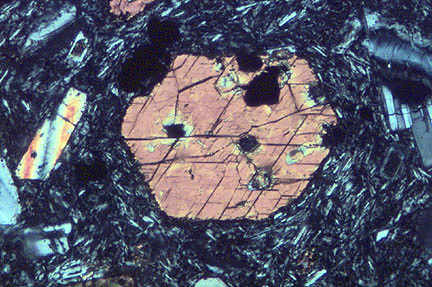
Hornblende
Thin Section Photomicrographs Of The Rotokawa Andesite A Download Scientific Diagram Earth And Space Science Earth From Space Andesite

Thin Section Description For Carbonate Grains Skeletal And Non Skeletal Matrix Cements And Pore Types For Environmen Too Thin Product Description Analysis

Snowball Garnets Are A Fascinating Structure And Beautiful Metamorphic Rocks Metamorphic Earth Photos

Calcarenite An Overview Sciencedirect Topics

Seismic Facies Analysis Seismic Geology Geophysics

Pressure Temperature Path Of Arquia Group Rocks Nw Colombia A Petrographic Analysis From Mineral Assemblage Assemblage Subduction Zone Metamorphic

Pin By Dana Lacavera On Nature S Treasures Minerals And Gemstones Rock Rocks And Minerals

Amphibole An Overview Sciencedirect Topics

Geol342 Sedimentation And Stratigraphy

Augite An Overview Sciencedirect Topics
