Transverse Section Of Brain

Brain Cross Section Diagram Cross Section Of The Human Brain Royalty Free Cliparts Vectors Brain Diagram Brain Pictures Human Brain

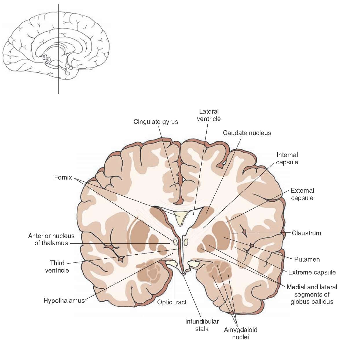
Frontal Section Taken Through The Level Of The Rostral Diencephalon Where The Thala Mus Is Not Present Note Again Th Gross Anatomy Brain Anatomy Brain Parts

Unlabeled Brain Diagram Thing That You Need To Learn Or Get Familiar With Your Brain System Human Anatomy Organs Le Brain Anatomy Brain Diagram Brain System
Cross Section Of The Brain Depicting The Positions Occupied By The Pulvinar Medial Geniculate In 2020 Nervous System Parts Central Nervous System Medical Illustration

Cross Section Of The Brain Depicting The Positions Occupied By The Pulvinar Medial Geniculate In 2020 Nervous System Parts Central Nervous System Medical Illustration

Horizontal Section Of The Brain Depicting 1 On One Side The Relationships Of The Internal Capsule To The In 2020 Nervous System Parts Internal Capsule Brain Anatomy
T2 weighting causes the nerve connections of white matter to appear gray and the congregations of neurons of gray matter to appear white while the cerebrospinal fluid csf appears light.
Transverse section of brain. This type of magnetic resonance imaging mri study looks at the brain with 20 transverse horizontal cuts beginning at the base and working to the top. Scroll through the images with detailed labeling using our interactive interface. 3 anatomical planes of human motion.
It is usually referred as the cut made between left and right. Lobes sulci and fissures of the cerebral hemispheres longitudinal fissure not pictured marieb hoehn human anatomy and physiology 9th ed. The structure whose name is clicked will be identified in the image by an arrow.
Bi 335 advanced human anatomy and physiology western oregon university figure 2. This tutorial has images in which the structures are labeled. Transverse section is a cut made in a plane that is made across the body of an animal a plant an organ or a tissue.
Smartdraw includes 1000s of professional healthcare and anatomy chart templates that you can modify and make your own. Axial mri atlas of the brain. Human movements are described in terms of three anatomical planes that run through the human body.
Free online atlas with a comprehensive series of t1 contrast enhanced t1 t2 t2 flair diffusion weighted axial images from a normal humain brain. You are to identify the structures by clicking on the name of the structure. Create healthcare diagrams like this example called the brain transverse section in minutes with smartdraw.
Sagittal coronal and transverse. Perfect for clinicians radiologists and residents reading brain mri studies. Each anatomical plane is governed by a set of positions and movements that help classify any physical activity.

Human Brain Cross Section Midsagittal View Cerebrum Frontal Parietal Temporal Occipital Lobes Cerebellum P Human Brain Anatomy Brain Pictures Brain Anatomy

1926 Human Anatomy Print Transverse Section Of Brain Etsy Anatomy Art Medical Anatomy Human Anatomy

Horizontal Section Depicting Internal Forebrain Structures After Parts Of The Cerebral Cortex Have Been Dis Nervous System Parts Internal Capsule Brain Anatomy

Transverse Sections Through The Brainstem Brain Anatomy Nervous Disease

Image Result For Transverse Section Of Medulla Oblongata Digestive System Anatomy Anatomy Medical

Transverse Section Of Brain Google Search Brain Stem Brain Anatomy Brain

Axial Section Through The Basal Ganglia The Left And Right Sections Are At Slightly Different Transverse Planes Anat Plexus Products Anatomy Drawing Images

The Brain Ion Cross Section Showing The Major Structures And Locations Of The Basal Nuclei Brain Diagram Brain Pictures Brain Structure

A Diagram Of A Cross Section Through The Upper Pons At The Level Of The Locus Brain Images Pons Brain Cranial Nerves

Transverse Section Of Medulla Oblongata Spinal Cord Anatomy Spinal Cord Craniosacral Therapy

Cross Sectional Diagram In Which The Principal Structures Of The Midbrain At The Level Of The Inferi Nervous System Parts Medical School Stuff Medical Pictures

Transverse Section Through Inferior Colliculus Medical School Studying Anatomy Medical Education

Transverse Section Through The Lower Part Of Pons Nervous System Anatomy Anatomy Gross Anatomy
