H E Staining Protocol Paraffin Sections

Haematoxylin Eosin H E Staining Protocols Online

Comparison Of Ihe And Traditional H E Staining A C Images Of Download Scientific Diagram

H E Staining Kit Hematoxylin And Eosin Ab245880 Abcam

Hematoxylin And Eosin H E Staining Of Liver Tissue A Control Download Scientific Diagram

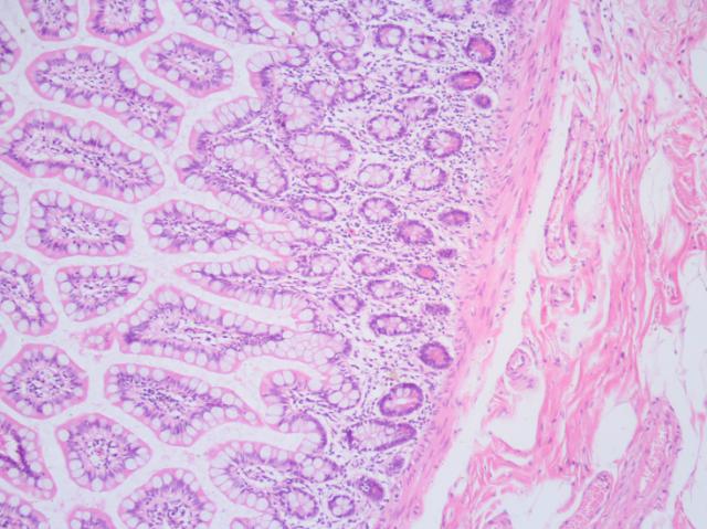
H E Staining Basics Troubleshooting Common H E Stain Problems Leica Biosystems
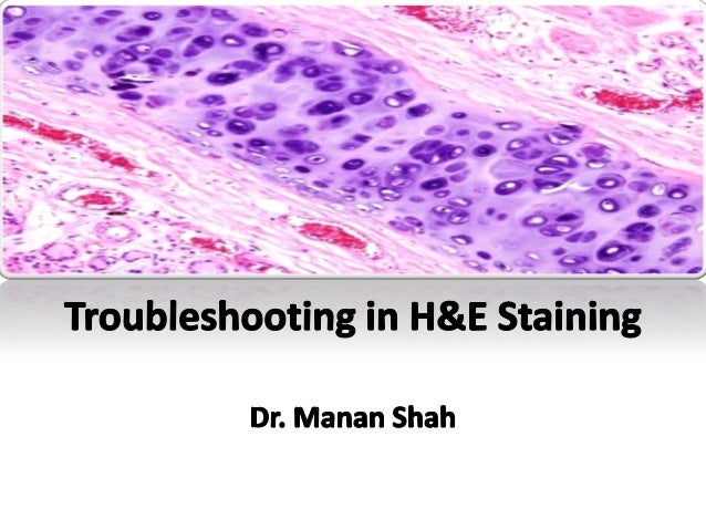
Troubleshooting In H E Staining

Ethanol 100 5 min.
H e staining protocol paraffin sections. Xylene desp 20 min. Ethanol 100 5 min. Use of very cold water slows down the.
Wash in running tap water for 5 minutes. 2 recommendations for deparaffinization. Deparaffinize sections 2 changes of xylene 10 minutes each.
The staining procedure for h e follows a basic protocol. Dewaxing dehydration hematoxylin differentiation bluing eosin dehydration clearing cover slipping. 3 x 3 xylene blot excess xylene before going into ethanol 3 x 3 100 ethanol 1 x 3 95 ethanol 1 x 3 80 ethanol 1 x 5 deionized h2o while sections are in water skim surface of hematoxalin with a kimwipe to remove oxidized particles.
1 p a g e h e staining paraffin embedded sections 1. Allow the slides to dry overnight and store slides at room temperature until ready for use. Use three changes of xylene 3.
Transfer the sections onto a superfrost plus slide. A few drops of strong ammonium hydroxide or of saturated aqueous lithium carbonate added immediately before use are sufficient for a 400 ml staining dish full of water. Section paraffin blocks at the desired thickness usually 4 5 µm on a microtome and float on a 40 c water bath containing distilled water.
If paraffin is not totally removed from tissue sections color intensity may be decreased or staining may be irregular spotty within the tissue section. Stain in harris hematoxylin solution for 8 minutes. Thus the stain discloses abundant structural information with specific functional implications.

H E Staining Of The Original Vaginal Tissue And 3d Cultures The Hnvec Download Scientific Diagram

Hematoxylin And Eosin H E Stain And Representative Distribution Of Download Scientific Diagram

Histology Of The Testes Of Treated And Untreated Group H E Staining Download Scientific Diagram

Figure Legends Figure 1 Histologic Examination Of Gingival Tissue Download Scientific Diagram

Hematoxylin And Eosin H E Stained Heart Sections From Mice Treated Download Scientific Diagram

Histological Analysis With H E Stain A H Hematoxylin Eosin Download Scientific Diagram

Hematoxylin And Eosin H E Staining Of Heart Aorta And Coronary Download Scientific Diagram

Cracks On Retinal Eye Of P0 Mice Post H E Staining
Hematoxylin And Eosin H E Histology Stain Control Histology Slides

Breast Tissue Samples Stained With Hematoxylin And Eosin H E Each Download Scientific Diagram
H E Staining Of Undecalcified And Decalcified Bone Tissue He Staining Download Scientific Diagram
Https Aasldpubs Onlinelibrary Wiley Com Doi Pdf 10 1002 Lt 23782

Hematoxylin And Eosin Staining Stain Glass Jars Jar
