Dicot Leaf Cross Section Domain

Cross Section Of Dicot Leaf Biology Worksheet Leaf Structure Things Under A Microscope

Leaf Anatomy Google Search Fancy Letters Late Birthday Wishes Late Birthday

Dicot Leaf Record Diagram Leaf Structure Things Under A Microscope Leaves

861d16c697765eb9e3c9a932f8d24b16 Jpg 1075 1072 Biology Plants Plant Science Botanical Science

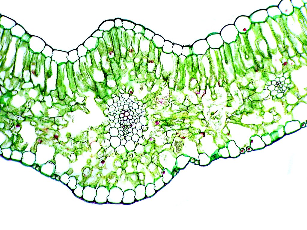
Dicot Leaf Cross Section Vascular Bundle Xylem Phloem Epidermis Palisade Mesophyll Spongy Mesoph Microscopic Photography Microscopic Cells Plant Tissue

Cross Section Of Dicot Leaf Cross Section Cross Blog Wallpaper

Monocot leaf shows parallel venation i e.
Dicot leaf cross section domain. However the main vein or midvein will always be seen in cross section see the ligustrummidvein shown below. F vein vascular bundle. Monocot leaves have bulliform cells on upper epidermis whereas in dicot leaves bulliform is absent.
J guard cell. What is monocot plant. E upper epidermis.
Learn vocabulary terms and more with flashcards games and other study tools. The cross section on the right is from a willow tree a dicot. G lower epidermis.
The palisade mesophyll produces carbohydrates by photosynthesis. K stoma function. If you wish to find out what these anatomical differences are type in monocot vs.
Cross section of a leaf. Anatomy of a dicot and monocot leaves leaves are very important vegetative organs because they are mainly concerned with photosynthesis and transpiration. Determine which characteristics belong to each category by writing an m or d in the table next to each.
Start studying monocot and dicot seed functions. Identify these leaves as belonging to a monocot or dicot. Both the leaf surfaces ventral and dorsal surface are similar because it has an equal number of stomata distribution and.

Dicot Leaf Cross Section Cross Section Cross Microscopic

Dicotyledon An Overview Sciencedirect Topics

Cross Section Of Dicot Leaf Leaves Blog Wallpaper Cross Section

Dicot Leaf Under Microscope Leaves Microscopic Anatomical

Pdf Three Dimensional Radiation Transfer Modeling In A Dicotyledon Leaf
Pdf How The Optical Properties Of Leaves Modify The Absorption And Scattering Of Energy And Enhance Leaf Functionality

Angiosperm And Gymnosperms Virtual Lab At College Of The Redwoods Studyblue

Monocot Dicot Leaves Slide Vwr

Pin By Hejer On Photosynthesis Leaf Structure Cell Structure Photosynthesis

Plants The Ohio State University At Lima

Angiosperm Morphology The Mesophytic Dicotyledonous Leaf Flickr
Internal Structure Of Root With Diagrams

Leaf Cross Section Dicot Vs Monocot Microscopic Photography Stem Structure Photo
